Israel Covid : Opinion: The COVID Vaccine Blood Libel Against Israel ... / Total and new cases, deaths per day, mortality and recovery rates, current active cases, recoveries, trends and timeline. . The only independent world health organization (who) recognized one stop platform for verified data and news. The country's coronavirus death toll remained unchanged at. Israel coronavirus update with statistics and graphs: דשבורד נתוני וירוס הקורונה בישראל מטעם משרד הבריאות הישראלי. Total and new cases, deaths per day, mortality and recovery rates, current active cases, recoveries, trends and timeline. The country's coronavirus death toll remained unchanged at. Total and new cases, deaths per day, mortality and recovery rates, current active cases, recoveries, trends and timeline. דשבורד נתוני וירוס הקורונה בישראל מטעם משרד הבריאות הישראלי. Israel coronavirus update with statistics and graphs: The only independent world health organization (who) recognized one s...
Dapatkan link
Facebook
X
Pinterest
Email
Aplikasi Lainnya
Anatomy Of Chest - Figure 7 From Relevant Surgical Anatomy Of The Chest Wall Semantic Scholar - Browse 2,533 female chest anatomy stock photos and images available, or start a new search to explore more stock photos and images.
Dapatkan link
Facebook
X
Pinterest
Email
Aplikasi Lainnya
-
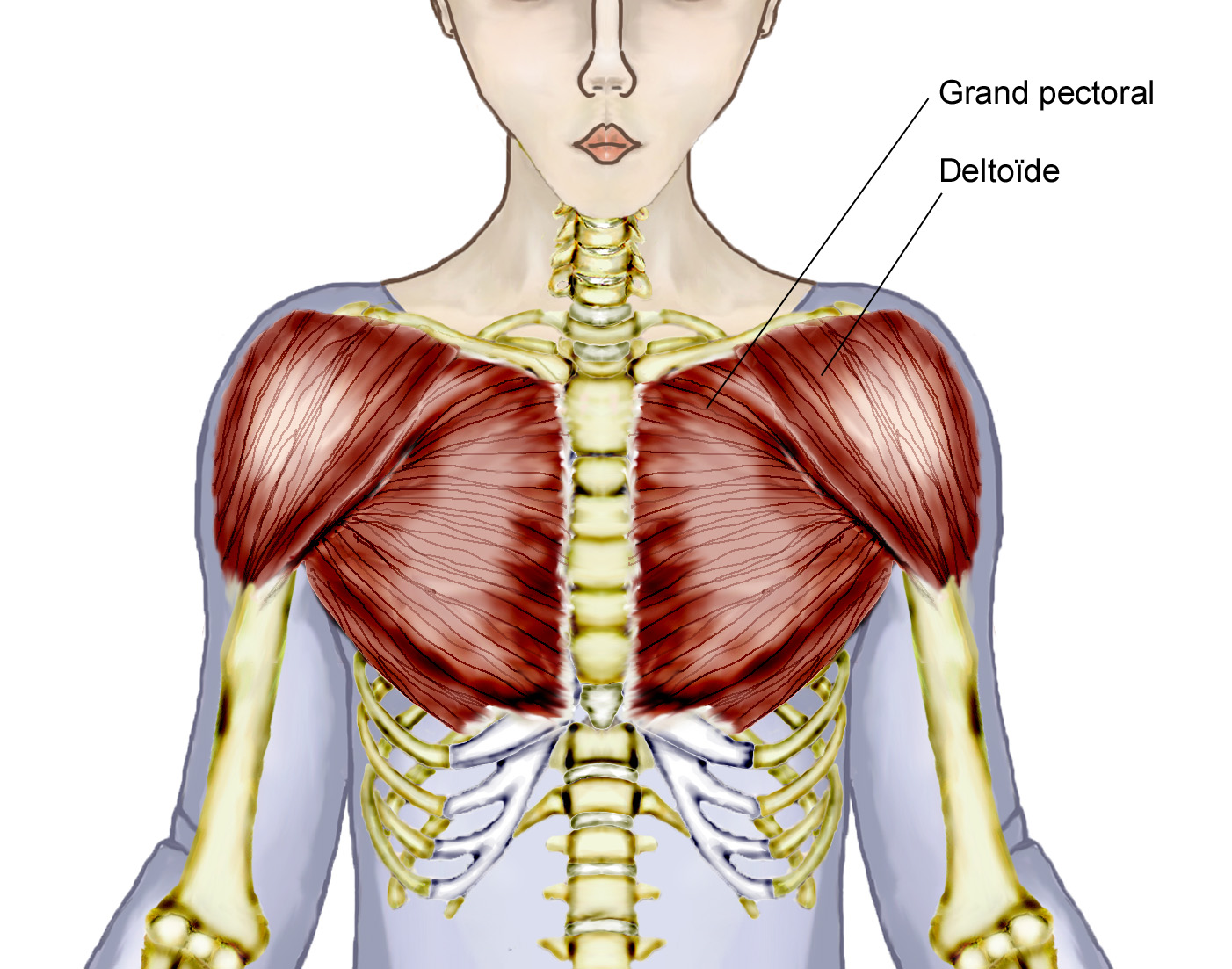
Anatomy Of Chest - Figure 7 From Relevant Surgical Anatomy Of The Chest Wall Semantic Scholar - Browse 2,533 female chest anatomy stock photos and images available, or start a new search to explore more stock photos and images.. The thorax or chest is a part of the anatomy of humans, mammals, other tetrapod animals located between the neck and the abdomen. The pec major) is the one that commands the most real estate. It is enclosed by the ribs, the vertebral column, and the sternum, or breastbone, and is separated from the abdominal cavity (the body's largest hollow space) by a muscular and membranous partition, the diaphragm. The palpable midline sternum is variable in size and shape; Of the two chest muscles, the pectoralis major (a.k.a.
The palpable midline sternum is variable in size and shape; This image added by admin. Anatomy of right side chest pain. The shape of the chest is often regarded as potential insight into a disease process, as in the case of barrel chest and respiratory dysfunction. It is enclosed by the ribs, the vertebral column, and the sternum, or breastbone, and is separated from the abdominal cavity (the body's largest hollow space) by a muscular and membranous partition, the diaphragm.
Diagram Female Chest Diagram Full Version Hd Quality Chest Diagram Diagramrt Ohimabrasserie It from www.modernheal.com Anatomy of the chest and shoulder, anatomy of the chest organs, anatomy of the chest wall, anatomy of the chest wall and pleura, anatomy of upper chest area, human. You can click the image to magnify if you cannot see clearly. Learn about each of these muscles, their locations, functional anatomy and exercises for them. This page provides an overview of the chest muscle group. Here, we break down the anatomy of your chest muscles. Fill out your shirt with a bigger, stronger, more powerful chest. It is made up of the manubrium superiorly, the body and the xiphisternum (figure 1).the manubrium has an upper central depression, the suprasternal notch. It provides protection to vital organs (eg, heart and major vessels, lungs, liver) and provides stability for movement.
The dominant muscle in the upper chest is the pectoralis major.
The chest wall is formed from the sternum anteriorly, 12 pairs of ribs, costal cartilages and intercostal muscles laterally, and the thoracic vertebrae posteriorly. Browse 2,533 female chest anatomy stock photos and images available, or start a new search to explore more stock photos and images. The chest is made up primarily of two muscles: You can click the image to magnify if you cannot see clearly. The epidermis is the outermost layer that provides a protective, waterproof seal over the body. We think this is the most useful anatomy picture that you need. The chest or thorax is the region between the neck and diaphragm that encloses organs, such as the heart, lungs, esophagus, trachea, and thoracic diaphragm. A typical heart is approximately the size of your fist: It is made up of the manubrium superiorly, the body and the xiphisternum (figure 1).the manubrium has an upper central depression, the suprasternal notch. Get the full built by science program: The pec major) is the one that commands the most real estate. Fill out your shirt with a bigger, stronger, more powerful chest. Of the two chest muscles, the pectoralis major (a.k.a.
This chapter is an abbreviated review of thoracic anatomy as seen on chest radiographs and computed tomography (ct) of the chest. About the 6th week, the somites differentiate into the sclerotomes and the dermatomyotomes. A good radiologist knows the anatomy because knowing where structures normally live and recognizing the location of an abnormality helps to make or narrow the differential diagnosis. Learn about each of these muscles, their locations, functional anatomy and exercises for them. Definition (nci_cdisc) the anterior side of the thorax from the neck to the abdomen.
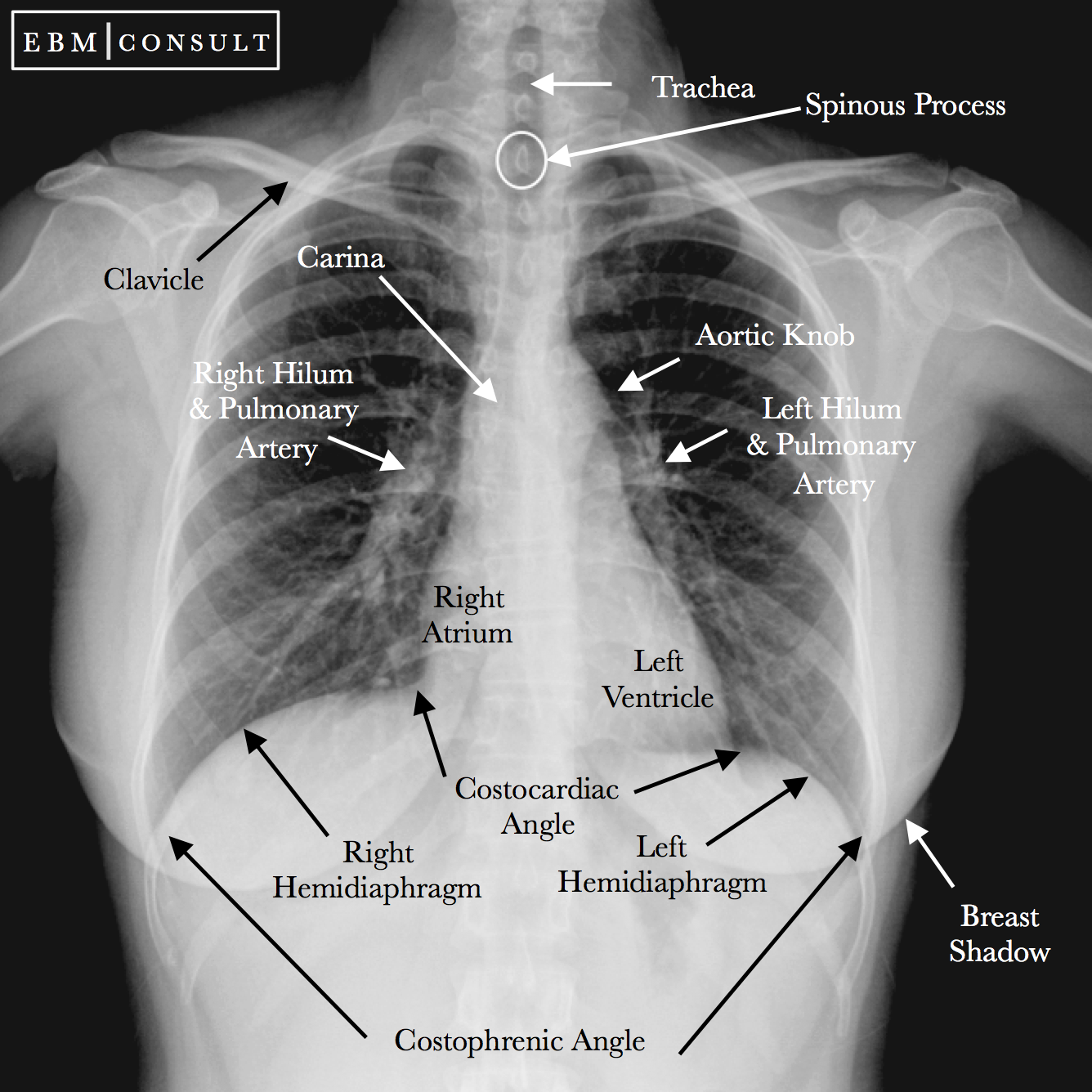
Radiology Chest Xray Normal from www.ebmconsult.com Plus, how to target each to make them bigger and stronger. Radiology basics of chest ct anatomy with annotated coronal images and scrollable axial images to help medical students and junior doctors learning anatomy. Thank you for visit anatomynote.com. A line is drawn from anterior surface of the body of 6th thoracic vertebrae passing through the apex of the heart up to anterior lower most part of diaphragm. Anatomy of right side chest pain. Having to do with the chest. Learn about each of these muscles, their locations, functional anatomy and exercises for them. However, the classical anatomical descriptions in textbooks make it difficult to gain full mastery of this subject, because the books usually deal with its elements separately.
First i'll do an intro to the different organs and structures in the chest, and then i'll go over some images showing their locations.
An overview of the anatomy visible in a transverse computed axial tomographical image of the thorax (and part of the abdomen) performed with intravenous cont. 12 cm (5 in) in length, 8 cm (3.5 in) wide, and 6 cm (2.5 in) in thickness. Fill out your shirt with a bigger, stronger, more powerful chest. This chapter is an abbreviated review of thoracic anatomy as seen on chest radiographs and computed tomography (ct) of the chest. Computed tomography (ct) of the chest can detect pathology that may not show up on a conventional chest radiograph(1). About the 6th week, the somites differentiate into the sclerotomes and the dermatomyotomes. Summary:for adequate treatment of patients with breast cancer, mastologists should have a complete understanding of the anatomy of the thoracic wall, axilla and breast. Here, we break down the anatomy of your chest muscles. Organs & structures of the chest heart. The epidermis is the outermost layer that provides a protective, waterproof seal over the body. The chest wall is comprised of skin, fat, muscles, and the thoracic skeleton. This page provides an overview of the chest muscle group. The chest anatomy includes the pectoralis major, pectoralis minor and the serratus anterior.
The epidermis is the outermost layer that provides a protective, waterproof seal over the body. Fill out your shirt with a bigger, stronger, more powerful chest. Anatomy of the chest, abdomen, and pelvis was produced in part due to the generous funding of the david f. Learn about each of these muscles, their locations, functional anatomy and exercises for them. Swensen fund for innovation in teaching.
Chest Anatomy from fpnotebook.com The anatomic illustrations are presented as… The epidermis is the outermost layer that provides a protective, waterproof seal over the body. Summary:for adequate treatment of patients with breast cancer, mastologists should have a complete understanding of the anatomy of the thoracic wall, axilla and breast. In insects, crustaceans, and the extinct trilobites, the thorax is one of the three main divisions of the creature's body, each of which is in turn composed of multiple segments. Here, we break down the anatomy of your chest muscles. This image added by admin. 12 cm (5 in) in length, 8 cm (3.5 in) wide, and 6 cm (2.5 in) in thickness. It provides protection to vital organs (eg, heart and major vessels, lungs, liver) and provides stability for movement.
However, the classical anatomical descriptions in textbooks make it difficult to gain full mastery of this subject, because the books usually deal with its elements separately.
The chest wall is comprised of skin, fat, muscles, and the thoracic skeleton. A line is drawn from anterior surface of the body of 6th thoracic vertebrae passing through the apex of the heart up to anterior lower most part of diaphragm. Learn about each of these muscles, their locations, functional anatomy and exercises for them. First i'll do an intro to the different organs and structures in the chest, and then i'll go over some images showing their locations. It is enclosed by the ribs, the vertebral column, and the sternum, or breastbone, and is separated from the abdominal cavity (the body's largest hollow space) by a muscular and membranous partition, the diaphragm. The shape of the chest is often regarded as potential insight into a disease process, as in the case of barrel chest and respiratory dysfunction. The chest or thorax is the region between the neck and diaphragm that encloses organs, such as the heart, lungs, esophagus, trachea, and thoracic diaphragm. You can click the image to magnify if you cannot see clearly. Definition (nci_cdisc) the anterior side of the thorax from the neck to the abdomen. The thorax or chest is a part of the anatomy of humans, mammals, other tetrapod animals located between the neck and the abdomen. Here's how science can help you grow! The chest is made up primarily of two muscles: Radiology basics of chest ct anatomy with annotated coronal images and scrollable axial images to help medical students and junior doctors learning anatomy.
Israel Covid : Opinion: The COVID Vaccine Blood Libel Against Israel ... / Total and new cases, deaths per day, mortality and recovery rates, current active cases, recoveries, trends and timeline. . The only independent world health organization (who) recognized one stop platform for verified data and news. The country's coronavirus death toll remained unchanged at. Israel coronavirus update with statistics and graphs: דשבורד נתוני וירוס הקורונה בישראל מטעם משרד הבריאות הישראלי. Total and new cases, deaths per day, mortality and recovery rates, current active cases, recoveries, trends and timeline. The country's coronavirus death toll remained unchanged at. Total and new cases, deaths per day, mortality and recovery rates, current active cases, recoveries, trends and timeline. דשבורד נתוני וירוס הקורונה בישראל מטעם משרד הבריאות הישראלי. Israel coronavirus update with statistics and graphs: The only independent world health organization (who) recognized one s...
Chelsea Vs Man City 2-1 - Oicjdr2 Awl0 M - Instant reactions after chelsea stun man city premier league. . A dress rehearsal for the champions league final. Thomas tuchel live press conference: Gilmour in, no mount or de bruyne as both managers make changes. While city and their fans were left to wonder what might have been, chelsea offered more evidence of the improvements they have made since thomas tuchel took charge at the start of the year. Chelsea fc player ratings vs man city: They deny city the chance to secure the premier league title today. 14 apr uefa champions league 14/04/2021. Report and highlights as kevin de bruyne and riyad mahrez settle thriller at the etihad in the champions' favour. Read about chelsea v man city in the premier league 2020/21 season, including lineups, stats and live blogs, on the official website of the premier league. A dress rehearsal for the champions league final. ...
Anderlecht Wallpaper Rsca Wallpaper - Breaking News Theprinceisback Youtube : On wallpaperset you can find only the best hd wallpapers and background pictures. . Digital, digital art, artwork, illustration, abstract, neon. Screen resolution can be found in the settings of your device, it would be right to. Preview the top 50 best wallpaper engine wallpapers of the year 2020! Groot assortiment merchandise voor de rsca fan. Don't use a title like i found a cool wallpaper of my favourite game or my first wallpaper, instead use a. Discover the magic of the internet at imgur, a community powered entertainment destination. Grootste fansite van rsca, beter bekend als royal sporting club anderlecht. The new rsca away uniform is in the. Download wallpapers anderlecht, 4k, logo, esl pro league, soccer, football club, belgium, grunge, rsc anderlecht, metal texture, anderlecht fc for desktop free. The best for your mobile device, desktop, smartphone, tablet, iphone, ipad and...


Komentar
Posting Komentar